Horse Hoof Anatomy: The Structures That Keep 1,200 lb on the Move
A horse can gallop at 35 mph, pivot on a dime, and land from a jump thanks to four hooves that convert concussion into forward motion. Each foot combines rigid horn, elastic pads, and a web of blood vessels that together act like a hydraulic shock absorber. Recognizing what sits where—and how the pieces interact—turns everyday picking, trimming, and shoeing into targeted preventive care rather than guess-and-check maintenance.
Why Knowing the Parts Matters
-
Guides trim angles that keep joints and tendons in biomechanical alignment
-
Pinpoints whether soreness begins in wall, sole, frog, or deeper laminar tissue
-
Reveals early signs of white-line disease, seedy toe, laminitis, or navicular stress before lameness appears
-
Informs shoeing decisions—steel, composite, glue-on, or barefoot—based on which structure needs protection
From Outside to Inside: Eight Key Structures
-
Hoof Wall – Three keratin layers (stratum externum, medium, internum). Bears 80 % of static weight, deflects rocks, and grows 6–10 mm per month—faster in summer, slower in drought.
-
White Line (Zona Alba) – Pale junction where inward-facing wall laminae lock into the sole’s terminal papillae. Functions as the capsule’s “glue strip”; stretching or dark lines here warn of laminar weakness or bacterial invasion.
-
Sole – Slightly concave, mildly flexible horn plate that shares load with the wall on soft ground. Healthy sole has 8–10 mm thickness at the tip of the frog and yields slightly under thumb pressure; a flat, thin sole transmits concussion directly to the coffin bone.
-
Frog – Wedge-shaped, elastin-rich pad with a high water content. Acts as a deformable hinge that cushions impact, grips slippery surfaces, and pumps venous blood back up the limb; a deep central sulcus or ragged edges suggest thrush or insufficient ground contact.
-
Collateral Grooves – Two channels flanking the frog. Their depth is a quick gauge of sole thickness (deep equals plenty, shallow warns of over-rasped horn). Packed debris here can foster white-line infection if not cleared.
-
Bars – Inward folds of the hoof wall extending from the heel forward along the collateral grooves. Stabilize the rear of the capsule and limit heel splay. Trimming bars too aggressively weakens heel support and encourages underrun heels.
-
Digital Cushion – Fibro-fatty wedge above the frog that compresses under load, dispersing shock to the lateral cartilages, and stores elastic energy for the next stride. Foals start with a mostly fatty cushion that remodels into tougher fibro-cartilage as mileage builds; stalled horses may lack this conditioning.
-
Lateral Cartilages & Coffin Bone (P3) – Flexible “wings” of cartilage rise from either side of the coffin bone. With each footfall they flex outward, widening the heels and aiding blood circulation. Ossified (mineralized) cartilages lose that flexion, reducing shock absorption and predisposing to sidebone-related lameness.
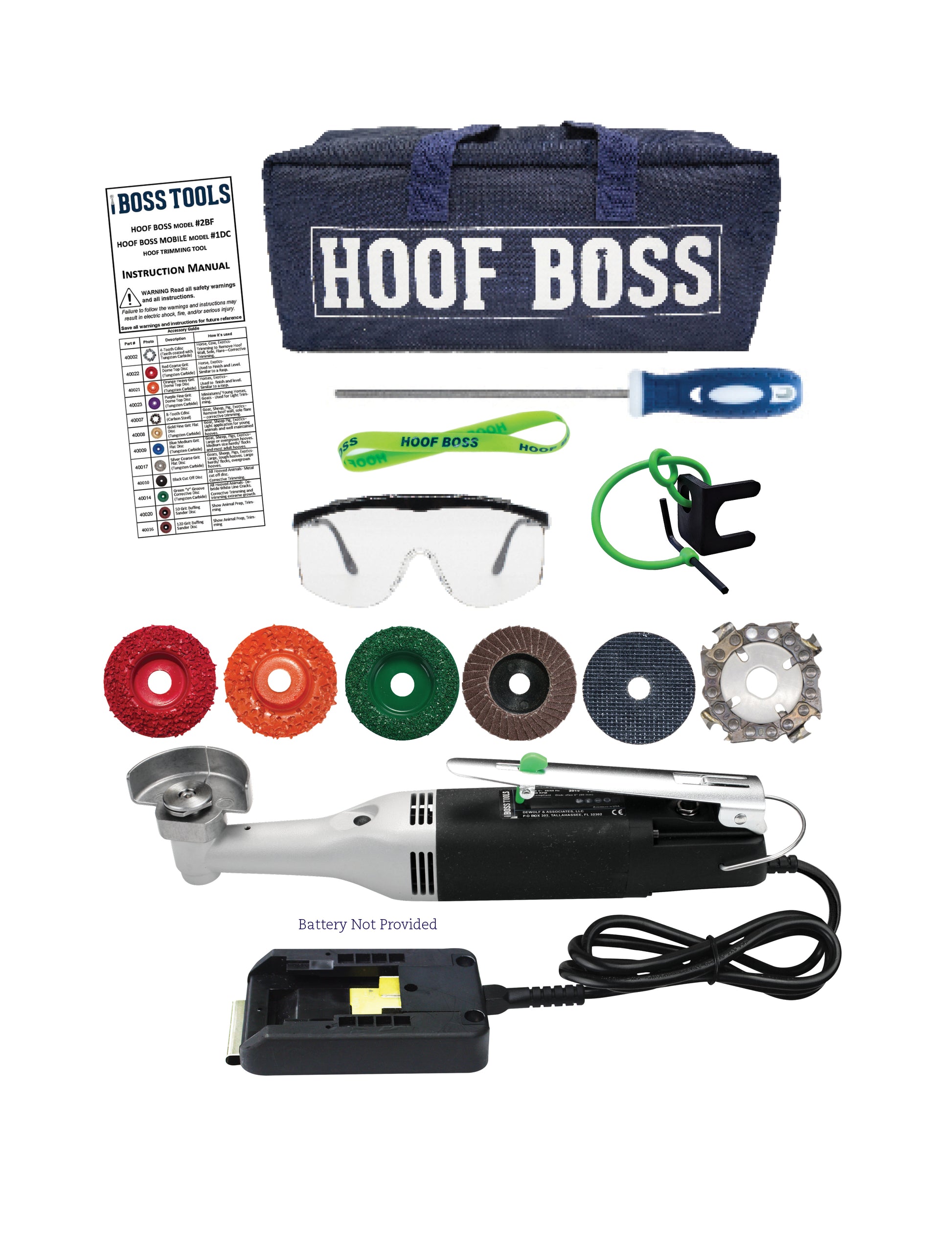
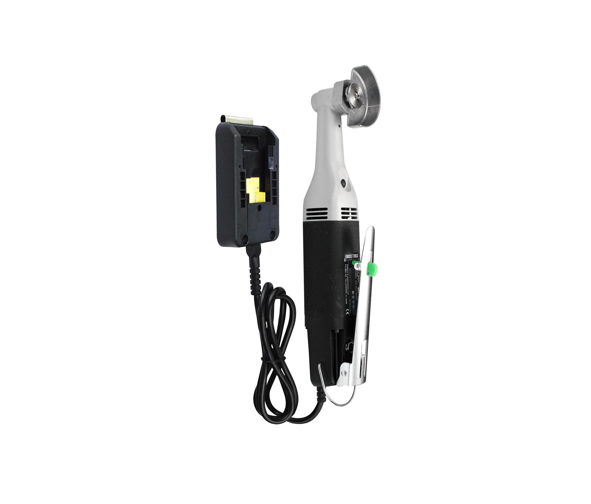
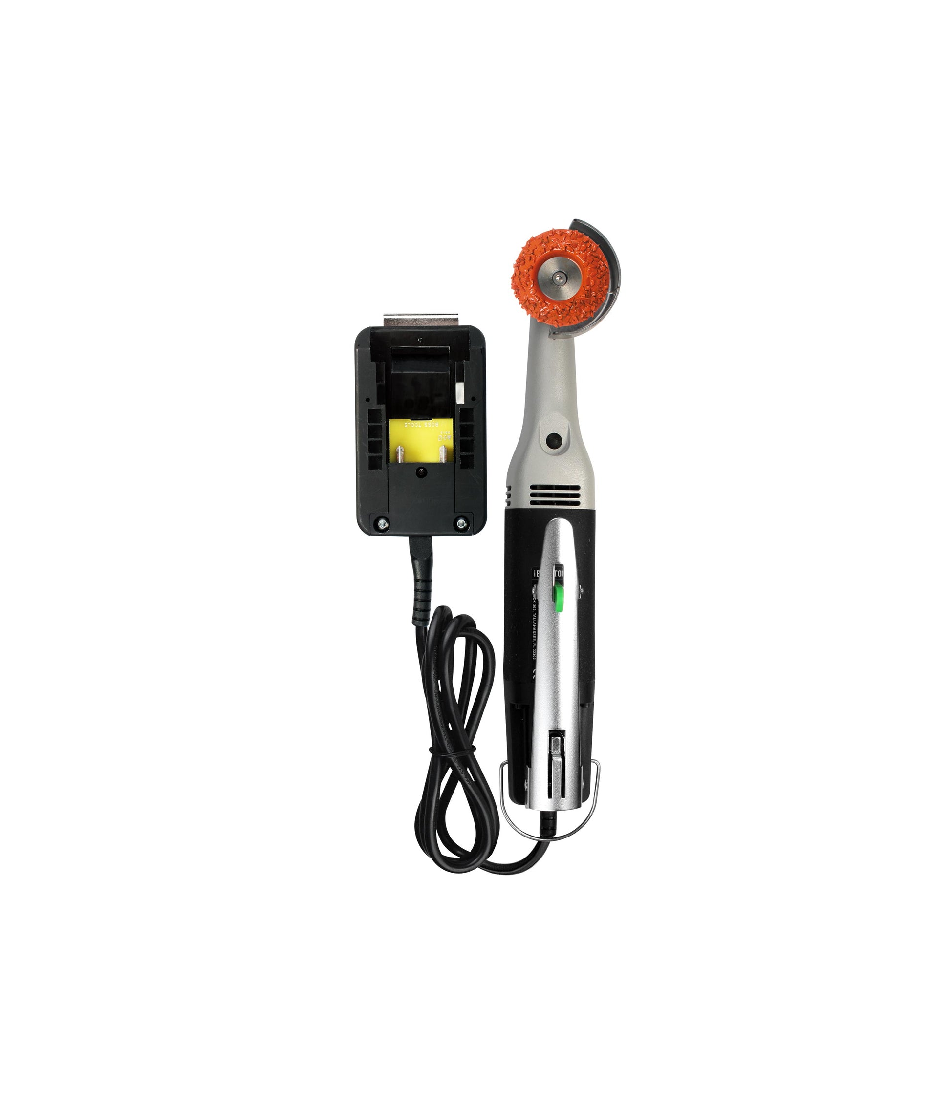
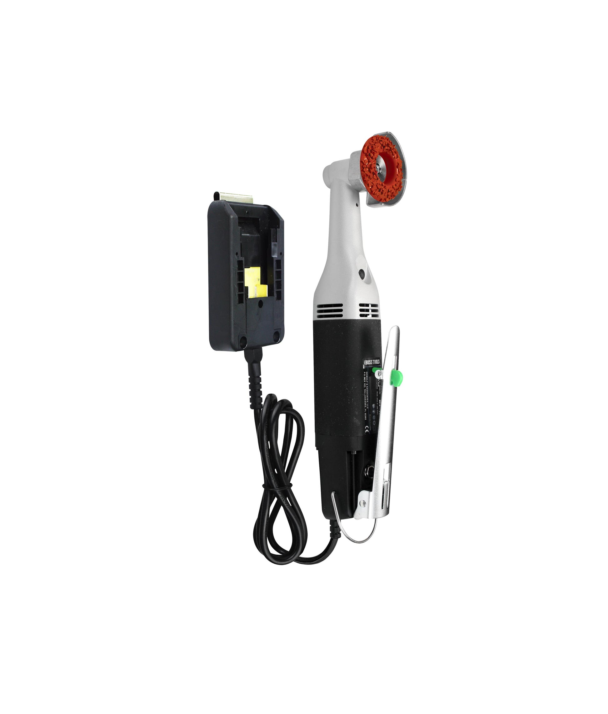
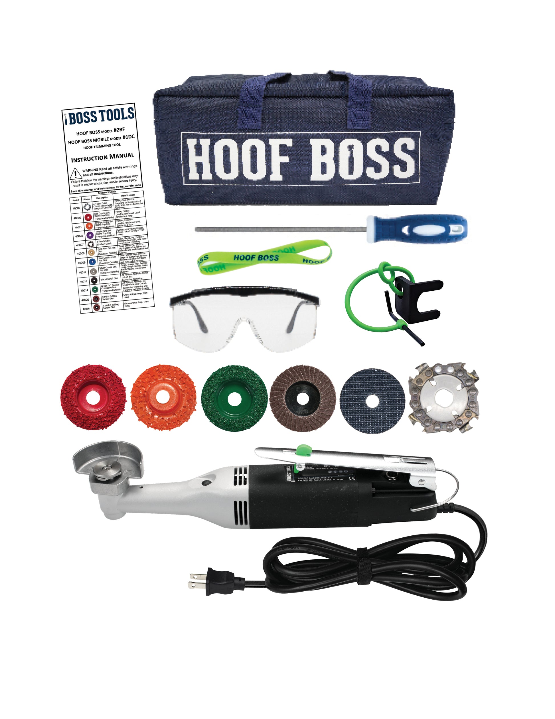
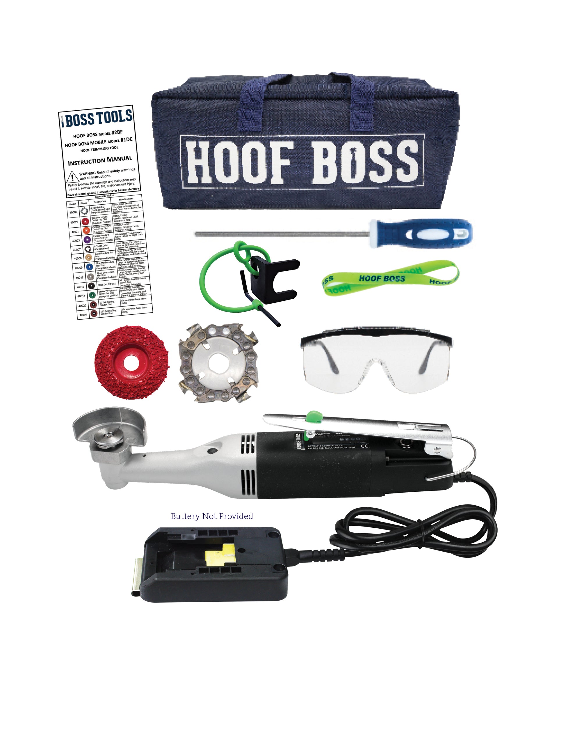
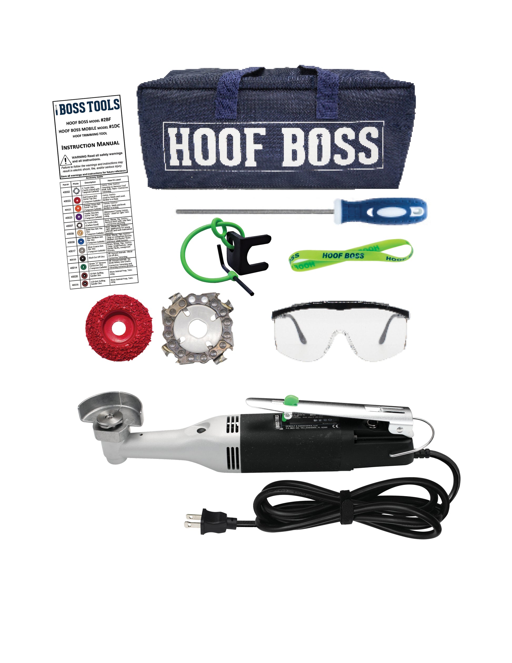
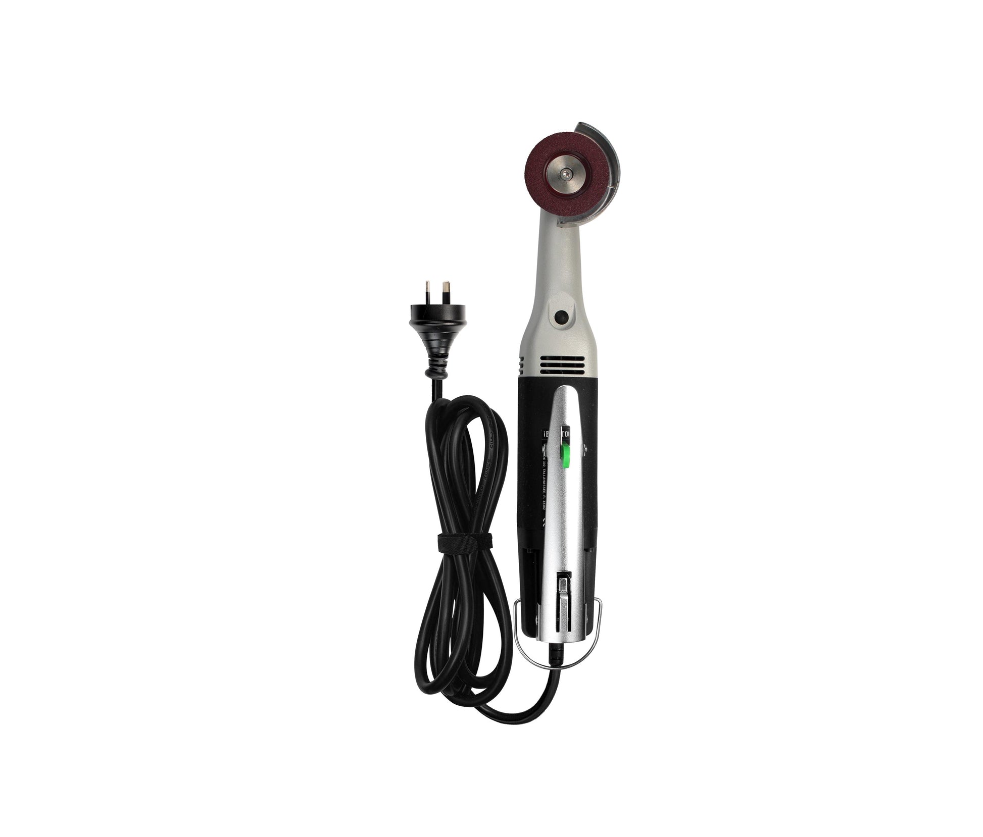
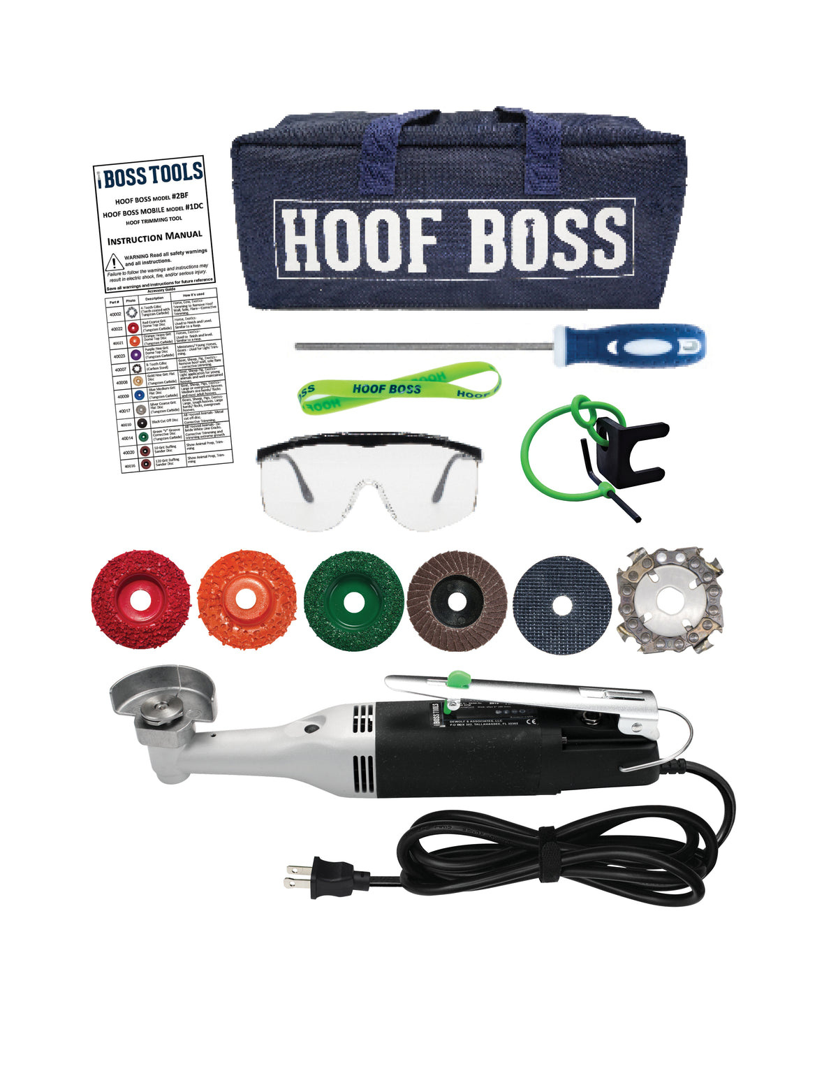
Between trims, horn continuously grows while environmental forces wear it away; balance is the goal. A wall that outruns sole leaves a perched hoof prone to cracks, while excess sole and frog can trap moisture and infection. Regular balancing—whether by rasp, knife, or a precision tool such as a variable-speed rotary trimmer with graded sanding discs—removes only the surplus horn and preserves each structure’s intended thickness.
Clean daily picking keeps collateral grooves open and allows the frog to engage the ground; adequate turnout on varied footing strengthens the digital cushion; a mineral-balanced diet supports dense horn growth; and correct shoe or boot selection protects any structure that is thin, bruised, or recovering from disease.
Knowledge of anatomy turns those management choices from rule-of-thumb into informed action, helping every stride stay elastic, confident, and pain-free.
Explore Hoof Boss Horse Hoof-Care Tools—equipment engineered for precise, anatomy-respectful maintenance.